
Search Product
How Does Glucose Cross Into The Cell
From a biophysical point of view, the entry of glucose into cells is not an arbitrary process. It relies primarily on a mechanism called ‘ease diffusion’ (Facilitated Diffusion), which is performed by a group of specialized carriers called glucose transporters (GLUTs).
Simply put, because glucose is a large molecule and polar, it cannot directly “walk through the wall” of the lipid bilayer of the cell membrane like oxygen. It must first bind to a specific GLUT protein on the cell surface. Once bound, this protein will undergo a conformational change (you can understand it as a physical deformation), and without directly consuming ATP, it will “transport” glucose into the membrane along the concentration gradient.
However, there are exceptions depending on the physiological needs of the organization. In tissues such as the intestine and kidney, glucose enters through secondary active transport (relying on SGLT protein), which is typical of “backwater” and is driven by the concentration difference of sodium ions. In muscle and fat cells, this process is completely “regulated” by insulin-only when instructed by insulin will the cell deploy the GLUT4 transporter to the cell membrane to receive glucose.
Why Glucose Can’t Drive In For A Long Time
To understand this transport mechanism, we must first look at the “wall”-the cell membrane. The cell membrane consists of a lipid bilayer, which is essentially a selective barrier. Its inner core is extremely hydrophobic.
According to our analysis of molecular properties, glucose wants to go in on its own and faces two hard injuries:
- Size: As a molecule, it is relatively too big.
- Polarity: Glucose is a polar molecule, which means that it is hydrophilic, but it is blocked by the lipophilic core of the cell membrane.
It is because of these properties that glucose cannot easily diffuse through the membrane like oxygen or carbon dioxide. It must rely on a “special doorway” or transport mechanism to bypass the limitations of the lipid bilayer.
Diffusion By GLUT Protein
In most cases, glucose enters the cell on the path of “easy diffusion. This process is entirely dependent on the family of glucose transporters (GLUTs). You can think of these transporters as specialized channels or vectors embedded in the cell membrane.
This workflow is very similar to the logic we use when designing precision valves:
- Binding: Glucose floating in the extracellular fluid binds to a specific site on the outward side of the GLUT protein.
- Conformational Change: Upon binding, the GLUT protein is immediately “deformed”. It reverses its orientation-it’s closed to the outside of the cell and open to the inside of the cell.
- Release: The glucose molecules are then released into the cytoplasm.
- Passive: The key point here is that the process itself does not require direct energy input (ATP). It operates entirely on a concentration gradient, allowing glucose to flow naturally from an area of high concentration (blood) to an area of low concentration (inside the cell).
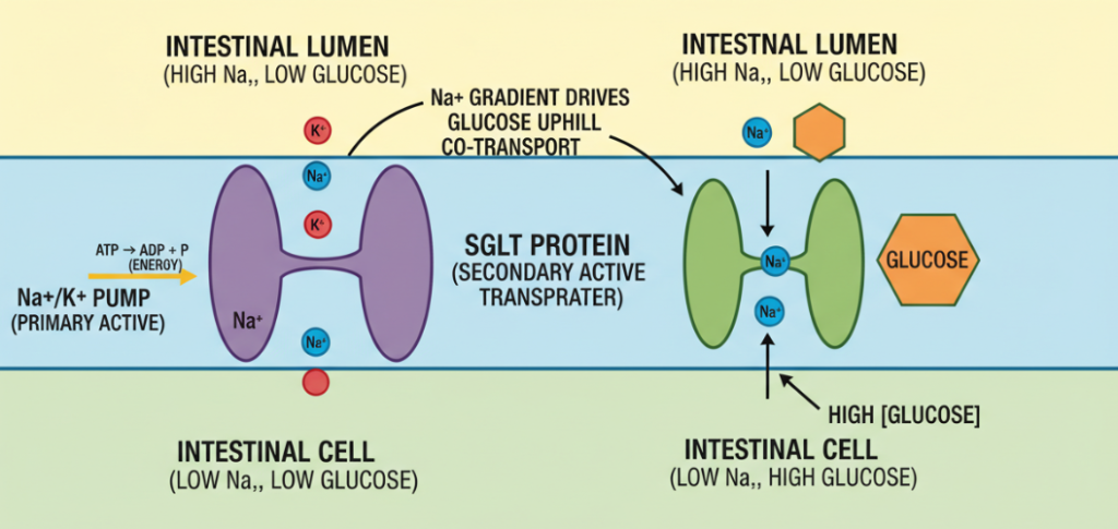
Secondary Active Transport
Although the efficiency of the diffusion is very high, it is along the concentration gradient. What if certain tissues (such as the intestines to absorb nutrients from food, or the kidneys to rescue sugar from urine back into the blood) need to continue to absorb glucose when the intracellular concentration of glucose is already high?
At this time, it is necessary to sodium-glucose coupled transporters (SGLTs) to come out for secondary active transport.
- Upstream (Against The Gradient): Unlike GLUTs, SGLTs can transport glucose against the concentration gradient.
- Sodium Driven: The system is powered by an electrochemical gradient of sodium ions. Cells consume energy through the sodium-potassium pump, forcibly maintaining an internal low sodium state. When external sodium ions rush into the cell to balance the concentration, it will “drag” glucose past the SGLT protein.
- Energy Consumption: Although ATP is not directly burned for transportation, in order to maintain the potential energy that allows sodium ions to rush in, the overall cell system consumes ATP indirectly.
Insulin Regulation And Translocation Of GLUT4
When doing research on metabolic products, we are most concerned about skeletal muscle and adipose tissue, because the glucose entry here is not “open all day long”, but is strictly regulated by hormones, especially insulin.
These cells depend on specific transporter: GLUT4.
At rest (when insulin levels are low), you won’t find GLUT4 on the cell surface. They are packed and stored in vesicles inside the cell, in a standby state.
The entire adjustment process is very precise:
- Insulin Signaling: When blood sugar rises, insulin binds to receptors on the surface of muscle or fat cells.
- Translocation (Translocation): This signal triggers a cascade of reactions that direct the GLUT4-loaded vesicles to move toward the cell membrane.
- Glucose Entry: The GLUT4 protein fuses with the cell membrane, transiently opening a channel for glucose by easy diffusion.
- Withdrawal: Once insulin levels drop, the GLUT4 transporter is recovered from the membrane and stored back inside the cell, closing the gate.
Author: Alan Reid
Hi, I am a cell biology researcher and science writer passionate about metabolic health. I specialize in simplifying complex physiological processes—like membrane transport and insulin signaling—to make them easy to understand for everyone.